Home

Korábban Néha szín ru pv ct száj Janice Izgatottnak lenni

Image | Radiopaedia.org

Chest CT with contrast, showing a low density enhanced mass (arrow) in... |  Download Scientific Diagram
Chest CT with contrast, showing a low density enhanced mass (arrow) in... | Download Scientific Diagram
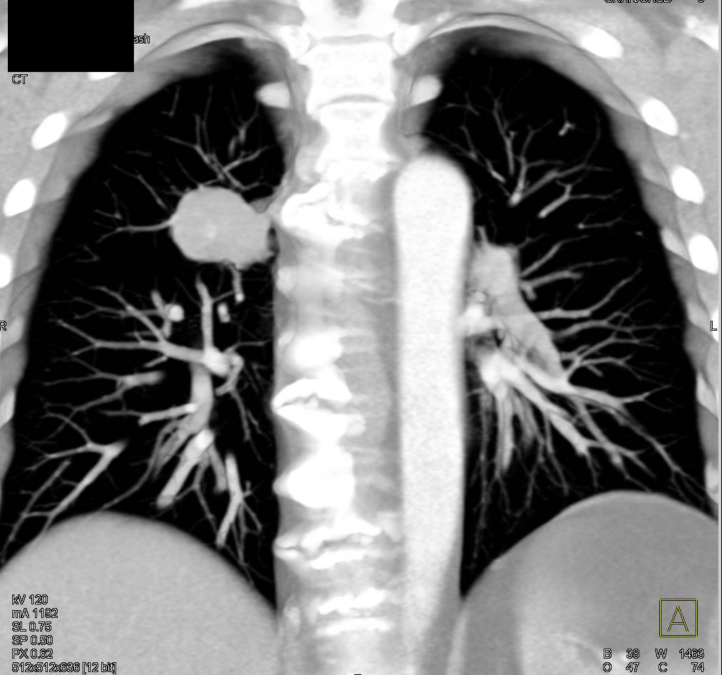
Pancreatic Cancer Encases the PV/SMV Junction with Lung Metastases As Well  - Pancreas Case Studies - CTisus CT Scanning
Pancreatic Cancer Encases the PV/SMV Junction with Lung Metastases As Well - Pancreas Case Studies - CTisus CT Scanning
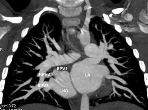
A Review on the Spectrum of Partial Anomalous Pulmonary Venous Connections:  The Added Value of Computed Tomography Imaging | Radiology and Medical  Diagnostic Imaging | Science Repository | Open Access
A Review on the Spectrum of Partial Anomalous Pulmonary Venous Connections: The Added Value of Computed Tomography Imaging | Radiology and Medical Diagnostic Imaging | Science Repository | Open Access

CT scan showed an unenhanced mass at the head of the pancreas (dotted... |  Download Scientific Diagram
CT scan showed an unenhanced mass at the head of the pancreas (dotted... | Download Scientific Diagram
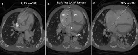
A Review on the Spectrum of Partial Anomalous Pulmonary Venous Connections:  The Added Value of Computed Tomography Imaging | Radiology and Medical  Diagnostic Imaging | Science Repository | Open Access
A Review on the Spectrum of Partial Anomalous Pulmonary Venous Connections: The Added Value of Computed Tomography Imaging | Radiology and Medical Diagnostic Imaging | Science Repository | Open Access

Solved QUESTION 8 1 points Save Answer Solve the formula for | Chegg.com

A) RUPV and RMPV are connected to the right superior caval vein.... |  Download Scientific Diagram
A) RUPV and RMPV are connected to the right superior caval vein.... | Download Scientific Diagram

Primary gastric sarcoma - recurrent | Radiology Case | Radiopaedia.org

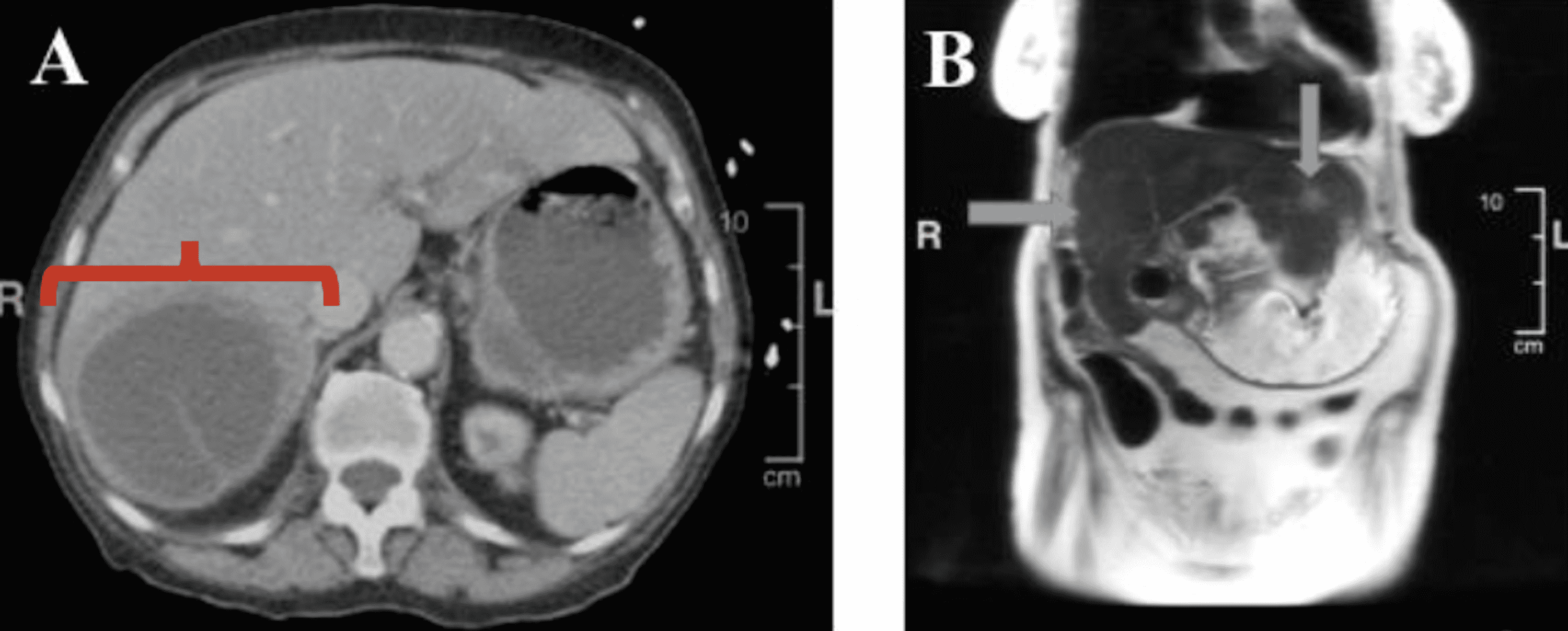
Cureus | Diagnosis of the Primary Tumor Site in the Case of Liver  Metastatic Carcinoma | Article
Cureus | Diagnosis of the Primary Tumor Site in the Case of Liver Metastatic Carcinoma | Article

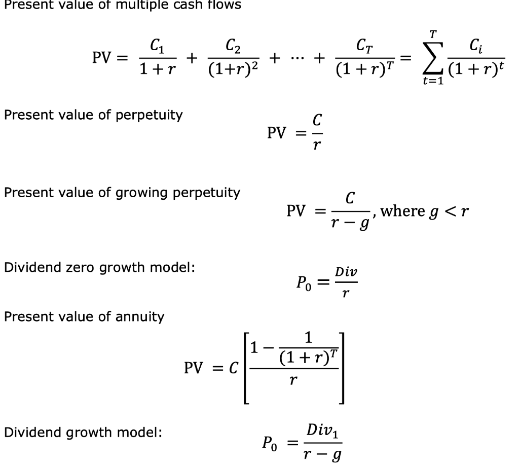
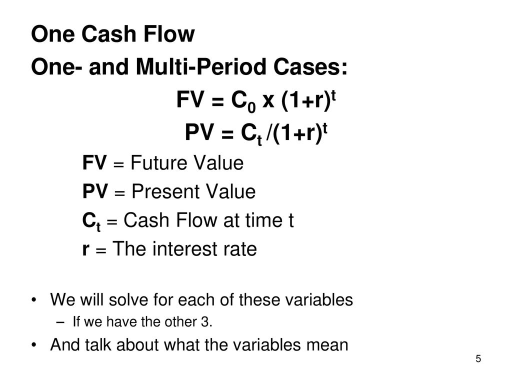
Solved Present value of multiple cash flows CT PV = C1 1+r | Chegg.com

Solved The present value of cash Ct received at a future | Chegg.com

Cardiac CT (A—axial section) showing both right upper lobe and left... |  Download Scientific Diagram
Cardiac CT (A—axial section) showing both right upper lobe and left... | Download Scientific Diagram
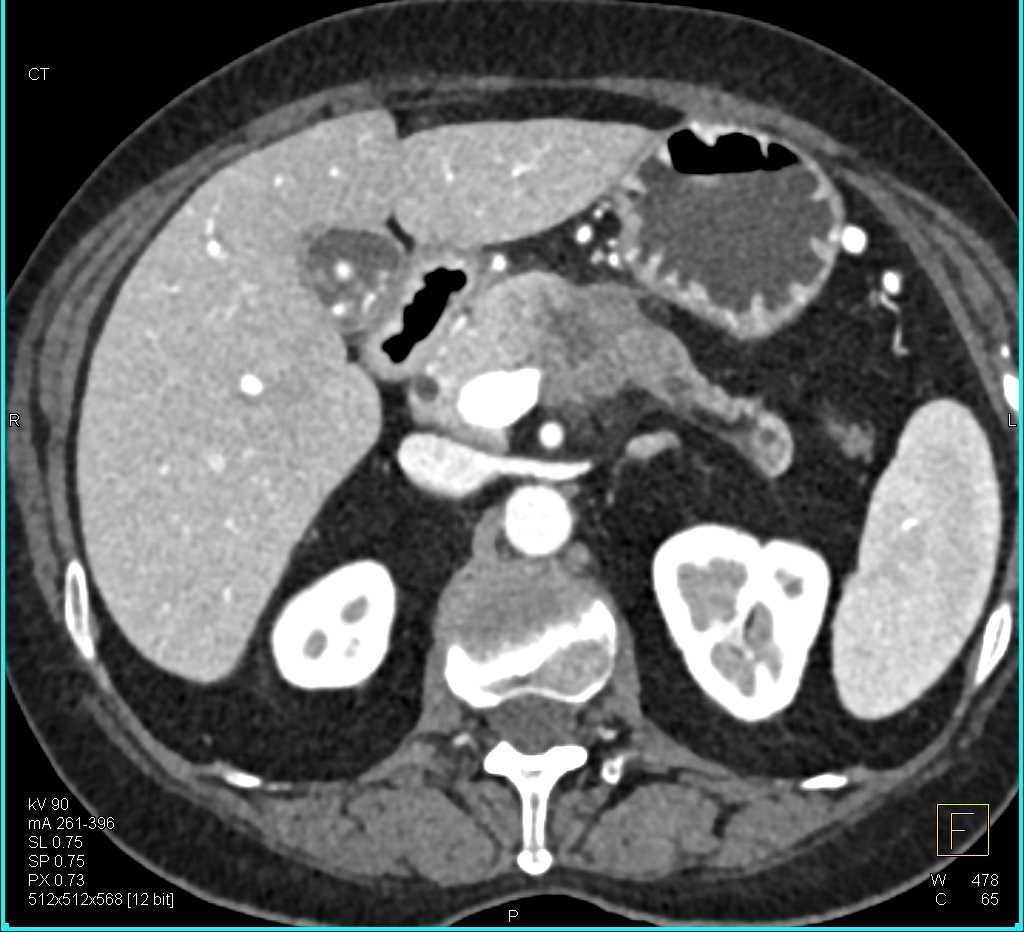
Carcinoma Body of the Pancreas Invades the PV-SMV Confluence - Pancreas  Case Studies - CTisus CT Scanning
Carcinoma Body of the Pancreas Invades the PV-SMV Confluence - Pancreas Case Studies - CTisus CT Scanning

A) Contrast enhanced chest computed tomography showed abnormal... |  Download Scientific Diagram
A) Contrast enhanced chest computed tomography showed abnormal... | Download Scientific Diagram

Pancreatic Cancer Encases the PV/SMV Junction - Pancreas Case Studies -  CTisus CT Scanning
Pancreatic Cancer Encases the PV/SMV Junction - Pancreas Case Studies - CTisus CT Scanning

Discounted Cash Flow Valuation and TVM - ppt download

Case 10 : A) CT 2D coronal image: Normal drainage of Right upper... |  Download Scientific Diagram
Case 10 : A) CT 2D coronal image: Normal drainage of Right upper... | Download Scientific Diagram

Primary Minimally Invasive Repair with Atriopericardial Anastomosis  Technique for Pulmonary Vein Stenosis after Catheter Ablation - Koichi  Inoue, Arudo Hiraoka, Genta Chikazawa, Taichi Sakaguchi, 2021
Primary Minimally Invasive Repair with Atriopericardial Anastomosis Technique for Pulmonary Vein Stenosis after Catheter Ablation - Koichi Inoue, Arudo Hiraoka, Genta Chikazawa, Taichi Sakaguchi, 2021
Solved Pr=c Cr 1 , roe, p=cut u = (6) t = ct.pt 2-ui- - 2 h | Chegg.com

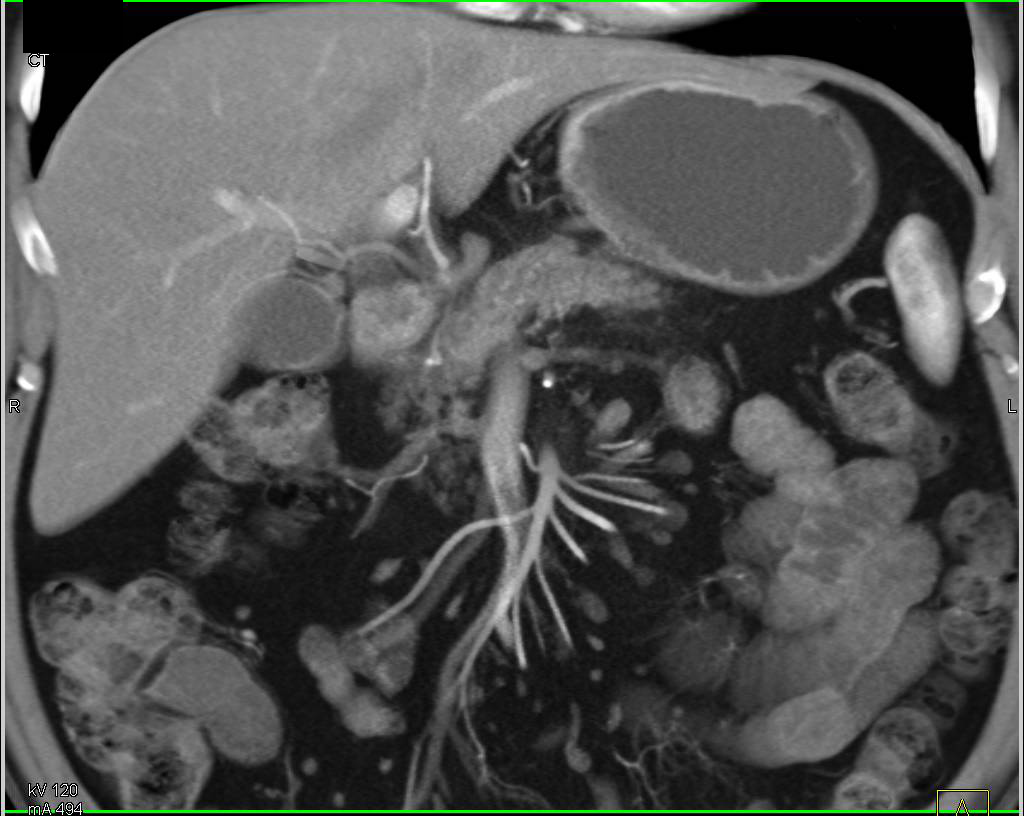
Pancreatic Cancer with Hepatic Artery and Portal Vein (PV) Involvement Best  Seen on 3D Images - Pancreas Case Studies - CTisus CT Scanning
Pancreatic Cancer with Hepatic Artery and Portal Vein (PV) Involvement Best Seen on 3D Images - Pancreas Case Studies - CTisus CT Scanning

Identifying Abdominal vessels on Axial CT scan - YouTube

Cantek CT-PV-S2M2812DIR-PS 1/2.7 2 Megapixel Auto Iris Day/Night CS-Mount,  2.8-12mm
Cantek CT-PV-S2M2812DIR-PS 1/2.7 2 Megapixel Auto Iris Day/Night CS-Mount, 2.8-12mm